What organs are made up nervous system
What organs are made up nervous system
Nervous system
Author: Jana Vasković MD • Reviewer: Nicola McLaren MSc
Last reviewed: August 15, 2022
Reading time: 20 minutes
The nervous system is a network of neurons whose main feature is to generate, modulate and transmit information between all the different parts of the human body. This property enables many important functions of the nervous system, such as regulation of vital body functions (heartbeat, breathing, digestion), sensation and body movements. Ultimately, the nervous system structures preside over everything that makes us human; our consciousness, cognition, behaviour and memories.
The nervous system consists of two divisions;
Understanding the nervous system requires knowledge of its various parts, so in this article you will learn about the nervous system breakdown and all its various divisions.
Cells of the nervous system
Two basic types of cells are present in the nervous system;
Neurons
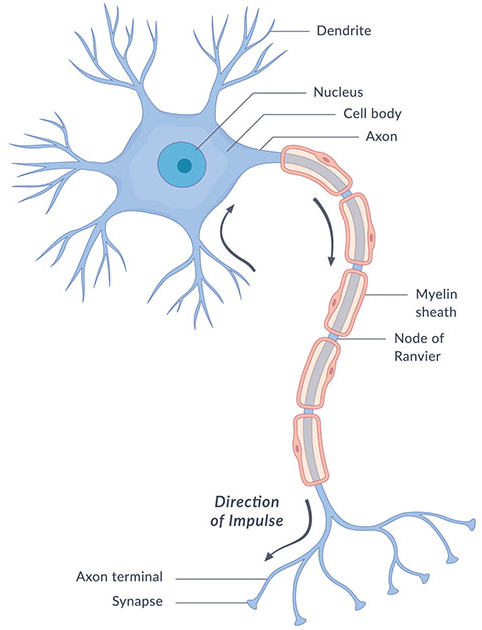
Neurons, or nerve cell, are the main structural and functional units of the nervous system. Every neuron consists of a body (soma) and a number of processes (neurites). The nerve cell body contains the cellular organelles and is where neural impulses (action potentials) are generated. The processes stem from the body, they connect neurons with each other and with other body cells, enabling the flow of neural impulses. There are two types of neural processes that differ in structure and function;
Every neuron has a single axon, while the number of dendrites varies. Based on that number, there are four structural types of neurons; multipolar, bipolar, pseudounipolar and unipolar.
Learn more about the neurons in our study unit:
How do neurons function?
The morphology of neurons makes them highly specialized to work with neural impulses; they generate, receive and send these impulses onto other neurons and non-neural tissues.
There are two types of neurons, named according to whether they send an electrical signal towards or away from the CNS;
The site where an axon connects to another cell to pass the neural impulse is called a synapse. The synapse doesn’t connect to the next cell directly. Instead, the impulse triggers the release of chemicals called neurotransmitters from the very end of an axon. These neurotransmitters bind to the effector cell’s membrane, causing biochemical events to occur within that cell according to the orders sent by the CNS.
Ready to reinforce your knowledge about the neurons? Try out our quiz below:
Glial cells
Glial cells, also called neuroglia or simply glia, are smaller non-excitatory cells that act to support neurons. They do not propagate action potentials. Instead, they myelinate neurons, maintain homeostatic balance, provide structural support, protection and nutrition for neurons throughout the nervous system.
This set of functions is provided for by four different types of glial cells;
Most axons are wrapped by a white insulating substance called the myelin sheath, produced by oligodendrocytes and Schwann cells. Myelin encloses an axon segmentally, leaving unmyelinated gaps between the segments called the nodes of Ranvier. The neural impulses propagate through the Ranvier nodes only, skipping the myelin sheath. This significantly increases the speed of neural impulse propagation.
White and gray matter
The white color of myelinated axons is distinguished from the gray colored neuronal bodies and dendrites. Based on this, nervous tissue is divided into white matter and gray matter, both of which has a specific distribution;
Master the histology of nervous tissue with our customizable quiz: We got you covered with neurons, nerves and ganglia!
Nervous system divisions
So nervous tissue, comprised of neurons and neuroglia, forms our nervous organs (e.g. the brain, nerves). These organs unite according to their common function, forming the evolutionary perfection that is our nervous system.
The nervous system (NS) is structurally broken down into two divisions;
Functionally, the PNS is further subdivided into two functional divisions;
They say that the nervous system is one of the hardest anatomy topic. But you’re in luck, as we’ve got a learning strategy for you to master neuroanatomy in a lot shorter time than you though you’ll need. Check out our quizzes and more for the nervous system anatomy practice!
Although divided structurally into central and peripheral parts, the nervous system divisions are actually interconnected with each other. Axon bundles pass impulses between the brain and spinal cord. These bundles within the CNS are called afferent and efferent neural pathways or tracts. Axons that extend from the CNS to connect with peripheral tissues belong to the PNS. Axons bundles within the PNS are called afferent and efferent peripheral nerves.
Central nervous system
The central nervous system (CNS) consists of the brain and spinal cord. These are found housed within the skull and vertebral column respectively.
The brain is made of four parts; cerebrum, diencephalon, cerebellum and brainstem. Together these parts process the incoming information from peripheral tissues and generate commands; telling the tissues how to respond and function. These commands tackle the most complex voluntary and involuntary human body functions, from breathing to thinking.
The spinal cord continues from the brainstem. It also has the ability to generate commands but for involuntary processes only, i.e. reflexes. However, its main function is to pass information between the CNS and periphery.
Learn more about the CNS anatomy here:
Peripheral nervous system
The PNS consists of 12 pairs of cranial nerves, 31 pairs of spinal nerves and a number of small neuronal clusters throughout the body called ganglia.
Peripheral nerves can be sensory (afferent), motor (efferent) or mixed (both). Depending on what structures they innervate, peripheral nerves can have the following modalities;
Cranial nerves
Cranial nerves are peripheral nerves that emerge from the cranial nerve nuclei of the brainstem and spinal cord. They innervate the head and neck. Cranial nerves are numbered one to twelve according to their order of exit through the skull fissures. Namely, they are: olfactory nerve (CN I), optic nerve (CN II), oculomotor nerve (CN III), trochlear nerve (CN IV), trigeminal nerve (CN V), abducens nerve (VI), facial nerve (VII), vestibulocochlear nerve (VIII), glossopharyngeal nerve (IX), vagus nerve (X), accessory nerve (XI), and hypoglossal nerve (XII). These nerves are motor (III, IV, VI, XI, and XII), sensory (I, II and VIII) or mixed (V, VII, IX, and X).
Among many strategies for learning cranial nerves anatomy, our experts have determined that one of the most efficient is through interactive learning. Check out Kenhub’s interactive cranial nerves quizzes and labeling exercises to cut your studying time in half.
Jump right into our cranial nerves quiz in multiple difficulty levels:
Or learn more about the cranial nerves in this study unit.
Spinal nerves
Spinal nervesemerge from the segments of the spinal cord. They are numbered according to their specific segment of origin. Hence, the 31 pairs of spinal nerves are divided into 8 cervical pairs, 12 thoracic pairs, 5 lumbar pairs, 5 sacral pairs, and 1 coccygeal spinal nerve. All spinal nerves are mixed, containing both sensory and motor fibers.
Spinal nerves innervate the entire body, with the exception of the head. They do so by either directly synapsing with their target organs or by interlacing with each other and forming plexuses. There are four major plexuses that supply the body regions;
Want to learn more about the spinal nerves and plexuses? Check out our resources.
Ganglia
Ganglia (sing. ganglion) are clusters of neuronal cell bodies outside of the CNS, meaning that they are the PNS equivalents to subcortical nuclei of the CNS. Ganglia can be sensory or visceral motor (autonomic) and their distribution in the body is clearly defined.
Dorsal root ganglia are clusters of sensory nerve cell bodies located adjacent to the spinal cord, They are a component of the posterior root of a spinal nerve.
Autonomic ganglia are either sympathetic or parasympathetic. Sympathetic ganglia are found in the thorax and abdomen, grouped into paravertebral and prevertebral ganglia. Paravertebral ganglia lie on either side of vertebral column (para- means beside), comprising two ganglionic chains that extend from the base of the skull to the coccyx, called sympathetic trunks. Prevertebral ganglia (collateral ganglia, preaortic ganglia) are found anterior to the vertebral column (pre- means in front of), closer to their target organ. They are further grouped according to which branch of abdominal aorta they surround; celiac, aorticorenal, superior and inferior mesenteric ganglia.
Parasympathetic ganglia are found in the head and pelvis. Ganglia in the head are associated with relevant cranial nerves and are the ciliary, pterygopalatine, otic and submandibular ganglia. Pelvic ganglia lie close to the reproductive organs comprising autonomic plexuses for innervation of pelvic viscera, such as prostatic and uterovaginal plexuses.
Find everything about ganglia needed for your neuroanatomy exam here.
Somatic nervous system
The somatic nervous system is the voluntary component of the peripheral nervous system. It consists of all the fibers within cranial and spinal nerves that enable us to perform voluntary body movements (efferent nerves) and feel sensation from the skin, muscles and joints (afferent nerves). Somatic sensation relates to touch, pressure, vibration, pain, temperature, stretch and position sense from these three types of structures.
Sensation from the glands, smooth and cardiac muscles is conveyed by the autonomic nerves.
Central Nervous System


The brain keeps the body in order. It helps to control all of the body systems and organs, keeping them working like they should. The brain also allows us to think, feel, remember and imagine. In general, the brain is what makes us behave as human beings.
The brain communicates with the rest of the body through the spinal cord and the nerves. They tell the brain what is going on in the body at all times. This system also gives instructions to all parts of the body about what to do and when to do it.
Spinal Cord 
Nerves divide many times as they leave the spinal cord so that they may reach all parts of the body. The thickest nerve is 1 inch thick and the thinnest is thinner than a human hair. Each nerve is a bundle of hundreds or thousands of neurons (nerve cells). The spinal cord runs down a tunnel of holes in your backbone or spine. The bones protect it from damage. The cord is a thick bundle of nerves, connecting your brain to the rest of your body.
Answer the questions / Ответьте на вопросы
What organs are made up nervous system?
What is the function of nervous system?
What parts of nervous system do you know?
What is the function of brain?
How does the brain communicate with the rest of the body?
Quiz
Instruction: Fill in the chart / Заполните таблицу
· Detect color and light
· Detects tastes: sweet, salty, sour and bitter
· Detects pain, pressure, heat and cold
| Sense | Organ | Job |
| Sight | ||
| Hearing | ||
| Smell | ||
| Taste | ||
| Touch |
Level B
Vocabulary
spinal cord – спинной мозг
to keep the body in order – поддерживать тело в порядке
to allow – позволять
forebrain – передний мозг
cerebral cortex – кора
to wire – связывать
Reading
The nervous system is made up of the brain, the spinal cord, and nerves. One of the most important systems in your body, the nervous system is your body’s control system. It sends, receives, and processes nerve impulses throughout the body. These nerve impulses tell your muscles and organs what to do and how to respond to the environment. There are three parts of your nervous system that work together: the central nervous system, the peripheral nervous system, and the autonomic nervous system.
The Nervous System
Hey, guys! Welcome to this Mometrix video over the nervous system.
All systems and parts of the body are important, but the nervous system is the system that has its fingers in everything your body does. It is the system that literally controls every process within your body.
The nervous system includes anything within the body that would allow you to sense; so, the brain, spine and spinal cord, all nerves, and any other sensory neurons.
Nervous System Function
The nervous system has four main roles or functions:
First, to collect and receive information internally and externally; so, from within the body and outside of the body.
Second, to process or derive meaning from the information collected and received.
Third, after the information has been processed, the nervous system forwards that information to the appropriate party; so, to the muscles, organs, or glands. This information sets them up to react how they need to.
Fourth, the nervous system dispatches the information to the regions of the brain that handle the processing of the information.
The nervous system, just like every other structure and makeup of our bodies, is very ordered and systematic. You can think of the nervous system as having two main divisions, and then several subdivisions that fit within those two divisions. The two divisions of the nervous system are the central nervous system (CNS) and the peripheral nervous system (PNS).
First, we will take a look at the central nervous system, and what exactly that includes.
Central Nervous System Function
The main function of the central nervous system is to incorporate the information it is getting and systemize the information so that it can queue a proper response.
The central nervous system is composed of the brain and the spinal cord. These two components encompass several other components. So, we will take a closer look at the brain first.
Regions of the Brain
The brain has six different regions: the cerebellum, cerebrum, medulla, brainstem, thalamus, and hypothalamus.
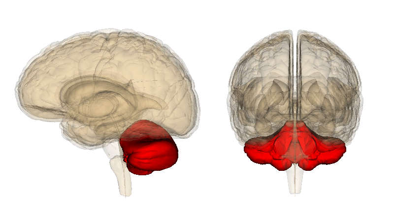
Cerebellum
The cerebellum sits right behind the upper portion of the brain stem (which is where the brain and spinal cord connect) and consists of two half-spheres (or hemispheres).
The cerebellum is the region of the brain that coordinates movement, and where various features of motor learning take place. In other words, the cerebellum is the part of your brain that allows for balance, coordination, speech, and posture, as well as things like muscle memory.
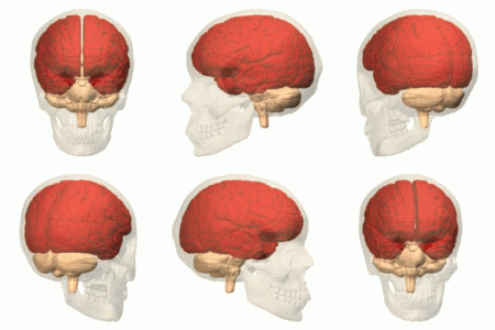
Cerebrum
The cerebrum sits in the top portion of the cranial cavity. The cerebrum is separated into a left and right hemisphere by a trench-like line called the longitudinal fissure. Now, perhaps counterintuitively, the left half of the cerebrum manages the right side of the body and the right half manages the left side of the body. However, even though there is a groove that distinctly separates the two halves of the cerebrum, there is a bundle of neural fibers, called the corpus callosum, that connects the two halves, and allows them to work together.
The cerebrum is made up of four different lobes: the frontal lobe, the temporal lobe, the parietal lobe, and the occipital lobe.
Frontal Lobe
The frontal lobe, like its name suggests, is at the front-most region of the brain, and, out of the four lobes, is the biggest.
The frontal lobe is split from the parietal and temporal lobe by fissures. The fissure that separates it from the parietal lobe is called the central sulcus, and the fissure that separates it from the temporal lobe, which is even deeper, is called the lateral sulcus. The frontal lobe contains the primary motor cortex. The primary central cortex (M1) is one of the main components of the brain included in the body’s motor function. The primary motor cortex initiates electrical signals and impulses that initiate body movement. The frontal lobe is also essential for problem solving, speech, and articulation, and it’s accountable for personality traits.
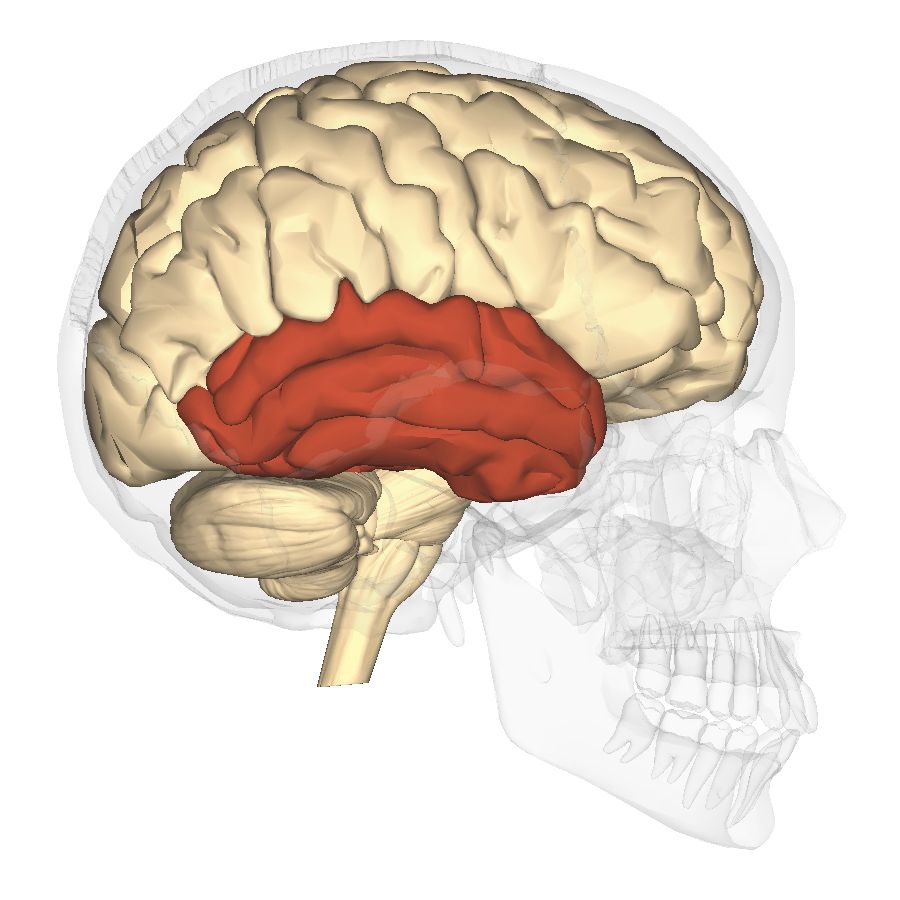
Temporal Lobe
You actually have two temporal lobes, one for both hemispheres. Your temporal lobes are located near your ears, and they mainly function in auditory processing. The temporal lobe taps into other roles as well, such as visual memory, your ability to learn language, and emotion association.
Parietal Lobe
The parietal lobe is set between the occipital lobe and the frontal lobe.
The parietal lobe has a really big job of having to process the information that it receives within milliseconds. The somatosensory cortex is located inside of the parietal lobe, and is crucial for processing the information from touch. The somatosensory cortex allows us to pinpoint the exact locality of touch perception, and helps to distinguish pain levels and temperature.
The parietal lobe is important for our depth of perception and communicates with other parts of the brain in order to perform other tasks.
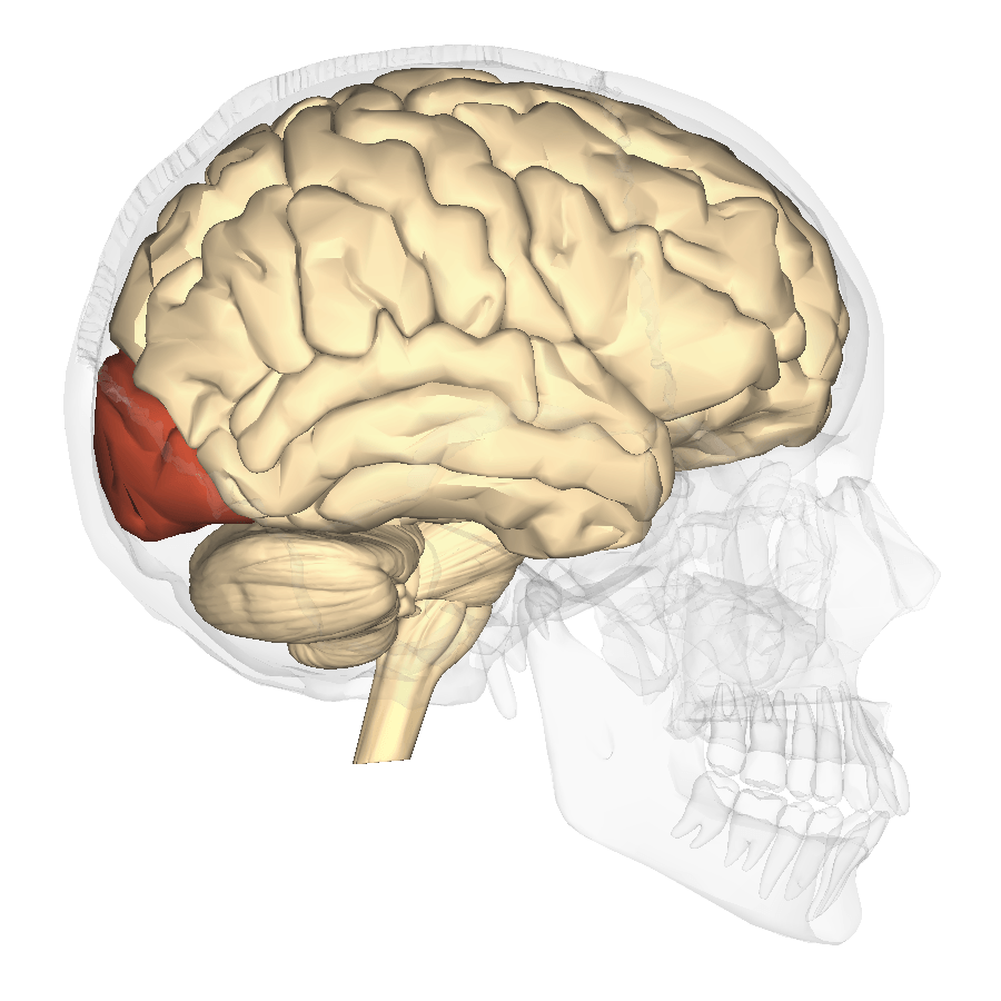
Occipital Lobe
The occipital lobe sits in the back part of the cortex.
The main function of the occipital lobe is to process visual information from the eyes and relay that information. The occipital lobe has to very quickly process that information, so that we are able to respond accordingly.
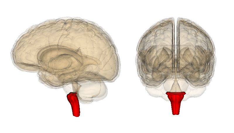
Medulla
That is an overview of the cerebrum, now let’s look at the medulla, more officially known as the medulla oblongata. The medulla is a section of your brainstem, and remember, your brainstem is where your brain and spinal cord connect. The medulla is one of the three parts of the brainstem.
The medulla has immediate control over numerous autonomic nervous system responses, it aids in the control of particular regions of the body, and it also plays a role in motor functions and outward motion. The medulla is very important, just like every other part of our brain. The medulla helps control breathing, and it controls our heart rate.
The medulla functions involuntarily. The medulla responds to various heart functions by dilating the blood flow so that there is a higher or lower amount of oxygen circulation, it can help by responding in fight or flight situations by switching digestion off or on, causes us to cough or sneeze to get rid of unwanted particles that get into your nasal cavity, and also controls vomiting or swallowing in order to dispel anything that may bring harm to you.
Brain Stem
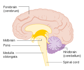
The fourth region of the brain that we will look at is the brain stem. The brain stem has three different parts: the medulla (which we just looked at), pons, and midbrain.
The pons sort of functions as a message headquarters for many other parts of the brain.
It aids in communication between brain sections, like the cerebrum and cerebellum. The pons is right above the medulla, and beneath the midbrain. So, it sits right in the middle. Many essential nerves stem from the pons. The vestibulocochlear nerve permits sound to proceed from our ears into our brain. The abducens nerve permits the eyes to move and look horizontally. The trigeminal nerve allows for us to experience feeling in the face, and the facial nerve allows for us to actually make facial expressions. The pons helps in the duties of the medulla, and it is linked to managing our sleep cycles.
Now, the midbrain is called the midbrain, because it sits between the forebrain and the hindbrain; but it’s important to note that out of the three parts of the brain stem it is not located in the middle. The midbrain functions, like the medulla, as this super message headquarters between the forebrain (or cerebral cortex) and the hindbrain (or the cerebellum and brainstem). The midbrain allows you to incorporate sensory data coming from your eyes and ears with data coming from your body’s muscle movements; helping it to adjust your movements based on important information.
All three parts of the brainstem function involuntarily.
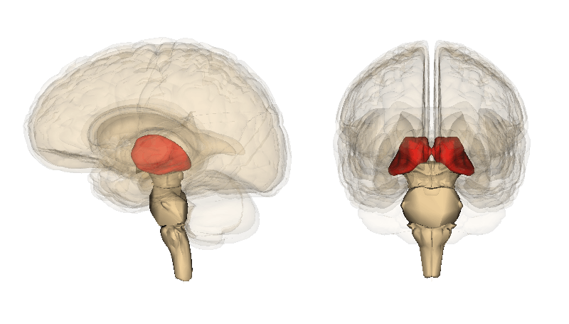
Thalamus
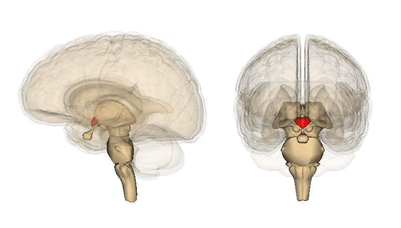
The thalamus is the fifth region of the brain. The thalamus sits right above the brainstem sandwiched between the midbrain and cerebral cortex.
The primary job of the thalamus is to inform the cerebral cortex of any sensory or motor stimuli. The thalamus also helps to control sleep, and aids in keeping us awake and alert. The thalamus, along with the hypothalamus, epithalamus, and subthalamus, is a part of the diencephalon, also sometimes called the interbrain. The diencephalon is the area where the vertebrate neural tube is located, which helps bring about the formation of the forebrain.
Hypothalamus
The sixth, and last, region of the brain is the hypothalamus. The hypothalamus is positioned below the thalamus and is the bottom part of the diencephalon.
One of the main functions of the hypothalamus is that it connects the nervous system to the endocrine system; which allows the hypothalamus to regulate our body’s temperature. The hypothalamus also aids in certain metabolic processes and helps to regulate the autonomic nervous system.
That was a closer look at the brain itself. The spinal cord is the other component that makes up the central nervous system. The spinal cord administers motor data coming from our brain to the appropriate parts of our body and helps to perform slight reflexes. It also administers sensory data directly from the peripheral nervous system to the brain. The spinal cord consists of 31 pairs of spinal nerves; because each of these nerves includes motor and sensory axons, they are referred to as mixed nerves.
So, that was the central nervous system. The second, main division of the nervous system is the peripheral nervous system.
Peripheral Nervous System
The peripheral nervous system is made up of nerves and what is called the ganglia.
Nerves are bundles of nerve fibers, also known as axons.
There are two types of nerves in the peripheral nervous system: spinal nerves and cranial nerves. There are 31 pairs of spinal nerves, and 12 pairs of cranial nerves. Ganglia refers to a cluster of neuron somas (or cell bodies). The individual cell body attaches to dendrites. The dendrites send information to neurons. The axons or nerve fibers then communicate that information to other neurons. It’s all a connected web. These nerves and ganglia are located around and outside of the brain and spinal cord.
The primary function of the peripheral nervous system is to link the central nervous system to the body’s organs and appendages, thus acting as the primary messenger between the brain, spinal cords, and everything else in the body. You can kind of think of the peripheral nervous system as the system that helps our brains and bodies to understand the world around us.
The peripheral nervous system has two subsystems: the somatic nervous system, and the autonomic nervous system.
Somatic Nervous System
The somatic nervous system is the part of the peripheral nervous system that is linked to voluntary bodily movements. It contains two parts, the sensory nervous system, as well as the somatosensory system, which is a part of the sensory nervous system.
The sensory nervous system is responsible for processing, as you might guess, sensory data. The most familiar sensory systems are the ones that control touch, taste, smell, hearing, and vision. The somatosensory system focuses on the conscious recognition of temperature, pain, touch, pressure, movement, position, and any sort of vibration. The somatosensory system communicates different sensations identified near the vicinity of the body via the spinal cord, brainstem, and up to the sensory cortex located in the parietal lobe. The sensory nervous system contains sensory neurons (or afferent neurons) and motor neurons (efferent neurons). Afferent neurons transmit stimuli to the central nervous system, as opposed to transmitting them from the CNS. Efferent neurons, in response to the afferent neurons, transmit signals from the central nervous system to the rest of the body. This transmission of stimuli through afferent neurons to the CNS, and then from the CNS to other parts of our body, allow for us to perform voluntary body movements as well as moderate involuntary reflex arcs.
Autonomic Nervous System
The autonomic nervous system is the other part of the peripheral nervous system that manages involuntary movements and functions—involuntary meaning that these movements happen without us be consciously in control of them happening. The autonomic nervous system also communicates information to our internal organs. There are two main divisions within the autonomic nervous system: the sympathetic nervous system, and the parasympathetic nervous system.
Sympathetic Nervous System
The sympathetic nervous system is the system that gets our body ready for the reaction that has been familiarized as the “fight or flight” response. When our body is being threatened or at risk for potential injury, this is the system that controls our response. The sympathetic nervous system causes our bodies to become keenly alert, speeds our heart rate, causes muscle contraction, and shuts off any function that is not essential for survival.
The overall functioning of the sympathetic nervous system is largely due to the neurotransmitter acetylcholine. This neurotransmitter binds to nicotinic receptors to trigger the release of norepinephrine which is produced by the adrenal gland to prepare our body and brain for action. Nicotinic receptors are the main receptors in the muscles for motor-neuron-to-muscle correspondence, that manage the contraction of muscles. The sympathetic nervous system originates in the thoracic and lumbar regions of the spinal cord.
Parasympathetic Nervous System
The parasympathetic nervous system, which is the other part of the autonomic nervous system, manages the body’s function when at rest. Rest and digest is the phrase given to reference the response of the parasympathetic nervous system. The parasympathetic nervous system works to conserve homeostasis within the body. It does the opposite of the sympathetic nervous system, and instead relaxes the body’s muscles, and slows our heart rate. Its job is to restore everything to a state of relaxation, and balance.
The parasympathetic nervous system increases bowel movements and urinary output. The neurotransmitter acetylcholine also plays a huge role in the parasympathetic nervous system by binding to muscarinic receptors, allowing the contraction of smooth muscles. This is what allows the parasympathetic to slow down the heart rate, regulate digestion, and relax muscles. The parasympathetic nervous system originates in the sacral portion of the spinal cord.
It’s important to note that both the central and peripheral nervous systems enlist the acetylcholine receptors, both nicotinic and muscarinic.
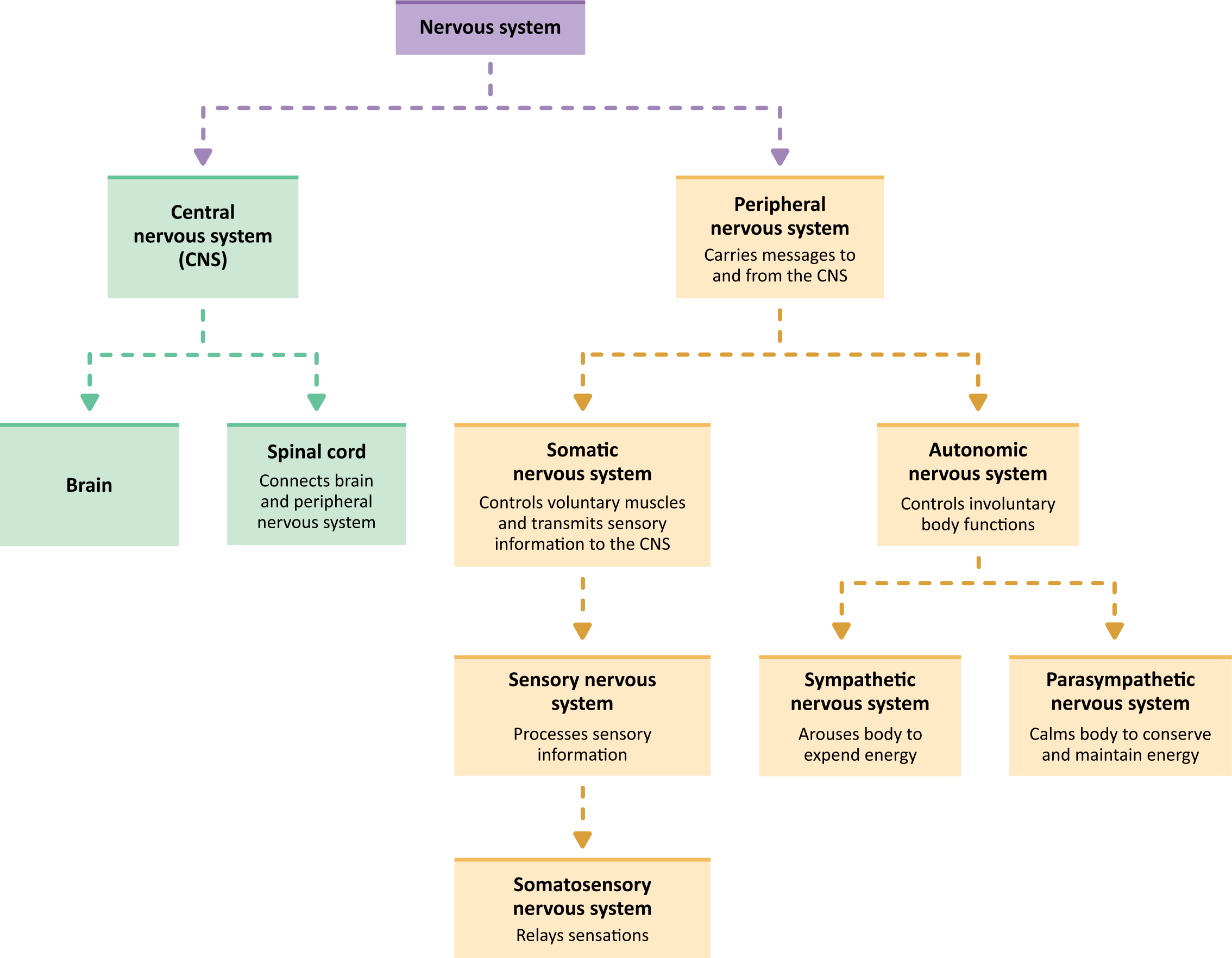
We just covered a lot of information. But, here is a very, very generalized diagram of the nervous system to help you remember the main parts.
You start with the nervous system itself. Then, you have the two main divisions: the central nervous system (which includes the brain and spinal cord), and the peripheral nervous system (which is made up of nerves and ganglia). The peripheral nervous system then has two subdivisions: the somatic nervous system (which controls voluntary movement), and the autonomic nervous system (which controls involuntary movement). Then, both the somatic and autonomic nervous systems contain two subdivisions. The somatic nervous system contains the sensory nervous system and the somatosensory system (which is a part of the sensory nervous system). And lastly, the autonomic nervous system contains the sympathetic nervous system (which controls our “fight or flight” response), and the parasympathetic nervous system which controls our “rest and digest” response).
If all of these systems seem pretty interconnected, the answer is yes.
I hope that this overview of the nervous system has been helpful to you!
What are the parts of the nervous system?
The nervous system has two main parts:
The nervous system transmits signals between the brain and the rest of the body, including internal organs. In this way, the nervous system’s activity controls the ability to move, breathe, see, think, and more. 1
The basic unit of the nervous system is a nerve cell, or neuron. The human brain contains about 100 billion neurons. A neuron has a cell body, which includes the cell nucleus, and special extensions called axons (pronounced AK-sonz) and dendrites (pronounced DEN-drahytz). Bundles of axons, called nerves, are found throughout the body. Axons and dendrites allow neurons to communicate, even across long distances.
Different types of neurons control or perform different activities. For instance, motor neurons transmit messages from the brain to the muscles to generate movement. Sensory neurons detect light, sound, odor, taste, pressure, and heat and send messages about those things to the brain. Other parts of the nervous system control involuntary processes. These include keeping a regular heartbeat, releasing hormones like adrenaline, opening the pupil in response to light, and regulating the digestive system.
When a neuron sends a message to another neuron, it sends an electrical signal down the length of its axon. At the end of the axon, the electrical signal changes to a chemical signal. The axon then releases the chemical signal with chemical messengers called neurotransmitters (pronounced noor-oh-TRANS-mit-erz) into the synapse (pronounced SIN-aps)—the space between the end of an axon and the tip of a dendrite from another neuron. The neurotransmitters move the signal through the synapse to the neighboring dendrite, which converts the chemical signal back into an electrical signal. The electrical signal then travels through the neuron and goes through the same conversion processes as it moves to neighboring neurons.
The nervous system also includes non-neuron cells, called glia (pronounced GLEE-uh). Glia perform many important functions that keep the nervous system working properly. For example, glia:
The brain is made up of many networks of communicating neurons and glia. These networks allow different parts of the brain to “talk” to each other and work together to control body functions, emotions, thinking, behavior, and other activities. 1,2,3
Тема2.7. The Nervous System/ Нервнаясистема
Level A
We learn the new words / Мыучимновыеслова
spinal cord – спинноймозг
to keep the body in order – поддерживатьтеловпорядке
to allow – позволять
cerebral cortex – кора
to wire – связывать
We read / Мычитаем
The nervous system is made up of the brain, the spinal cord, and nerves. One of the most important systems in your body, the nervous system is your body’s control system. It sends, receives, and processes nerve impulses throughout the body. These nerve impulses tell your muscles and organs what to do and how to respond to the environment. There are three parts of your nervous system that work together: the central nervous system, the peripheral nervous system, and the autonomic nervous system.
Central Nervous System

The brain keeps the body in order. It helps to control all of the body systems and organs, keeping them working like they should. The brain also allows us to think, feel, remember and imagine. In general, the brain is what makes us behave as human beings.
The brain communicates with the rest of the body through the spinal cord and the nerves. They tell the brain what is going on in the body at all times. This system also gives instructions to all parts of the body about what to do and when to do it.
Spinal Cord 
Nerves divide many times as they leave the spinal cord so that they may reach all parts of the body. The thickest nerve is 1 inch thick and the thinnest is thinner than a human hair. Each nerve is a bundle of hundreds or thousands of neurons (nerve cells). The spinal cord runs down a tunnel of holes in your backbone or spine. The bones protect it from damage. The cord is a thick bundle of nerves, connecting your brain to the rest of your body.
Answer the questions / Ответьтенавопросы
What organs are made up nervous system?
What is the function of nervous system?
What parts of nervous system do you know?
What is the function of brain?
How does the brain communicate with the rest of the body?
Quiz
Instruction: Fill in the chart / Заполнитетаблицу
· Detect color and light
· Detects tastes: sweet, salty, sour and bitter
· Detects pain, pressure, heat and cold
| Sense | Organ | J ob |
| Sight | ||
| Hearing | ||
| Smell | ||
| Taste | ||
| Touch |
LevelB
Vocabulary
spinalcord – спинной мозг
tokeepthebodyinorder – поддерживать тело в порядке
forebrain – передний мозг
Reading
The nervous system is made up of the brain, the spinal cord, and nerves. One of the most important systems in your body, the nervous system is your body’s control system. It sends, receives, and processes nerve impulses throughout the body. These nerve impulses tell your muscles and organs what to do and how to respond to the environment. There are three parts of your nervous system that work together: the central nervous system, the peripheral nervous system, and the autonomic nervous system.
Central Nervous System

The brain keeps the body in order. It helps to control all of the body systems and organs, keeping them working like they should. The brain also allows us to think, feel, remember and imagine. In general, the brain is what makes us behave as human beings.
The brain communicates with the rest of the body through the spinal cord and the nerves. They tell the brain what is going on in the body at all times. This system also gives instructions to all parts of the body about what to do and when to do it.
Spinal Cord 
Nerves divide many times as they leave the spinal cord so that they may reach all parts of the body. The thickest nerve is 1 inch thick and the thinnest is thinner than a human hair. Each nerve is a bundle of hundreds or thousands of neurons (nerve cells). The spinal cord runs down a tunnel of holes in your backbone or spine. The bones protect it from damage. The cord is a thick bundle of nerves, connecting your brain to the rest of your body.
Central Nervous
«Senses»
Senses
The cerebrum is part of the forebrain. The cerebral cortex is the outer layer of the cerebrum. Certain areas of the cerebral cortex are involved with certain functions.

The Peripheral Nervous System

Answer the questions
What organs are made up nervous system?
What is the function of nervous system?
What parts of nervous system do you know?
What is the function of brain?
How does the brain communicate with the rest of the body?
What senses do you know?
What is the function of neuron?
Quiz
Instruction: Fill the blank
· Detect color and light
· Detects tastes: sweet, salty, sour and bitter
:background_color(FFFFFF):format(jpeg)


